Boost precision across readers with AI that highlights 11 major thoracic abnormalities with 95-100% AUC* (and supports tuberculosis screening), compares priors, and supports earlier diagnosis.
*Clinical Evaluation Report of Lunit INSIGHT CXR_Rev.9
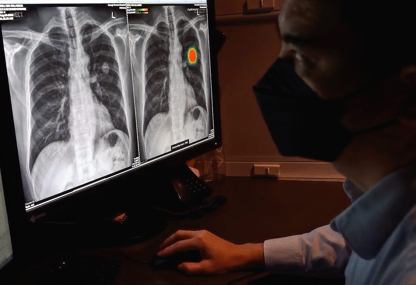
Lunit INSIGHT CXR is a clinically validated X-ray AI solution designed to support radiologists.
It helps identify both common and complex pathologies with greater speed and precision.
Powered by Lunit’s DualScan System, it combines two chest AI engines—one for abnormality detection and one for flagging normal cases—to help radiologists quickly identify suspicious reads and reduce volume in high-throughput environments.
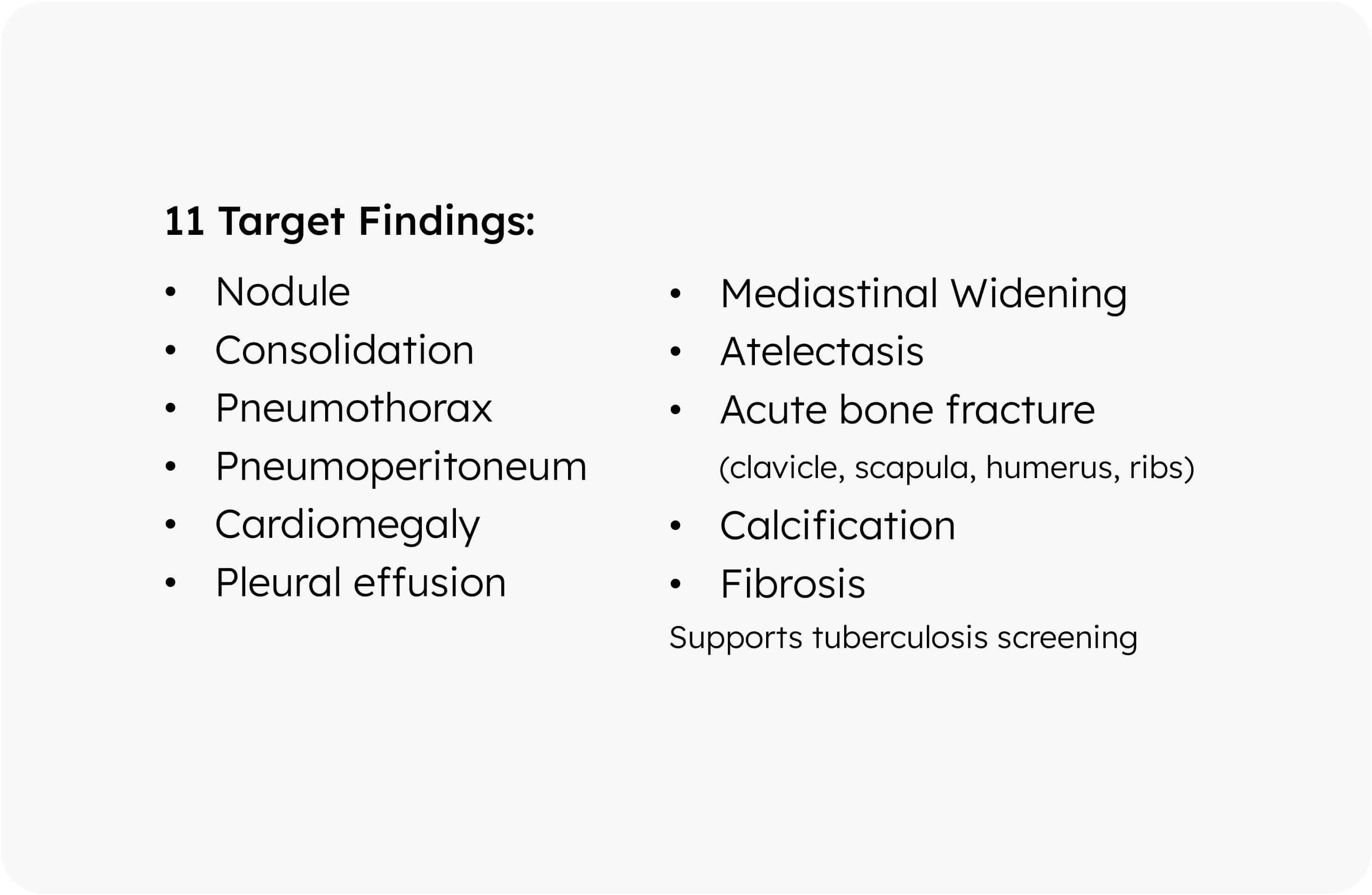
Lunit INSIGHT CXR achieves up to 100% accuracy* in detecting 11 major thoracic abnormalities—including acute bone fractures, nodules, pneumothorax, consolidation, and pleural effusion. Its performance has been independently validated on a training dataset of 1 million images.
*Clinical Evaluation Report of Lunit INSIGHT CXR_Rev.9
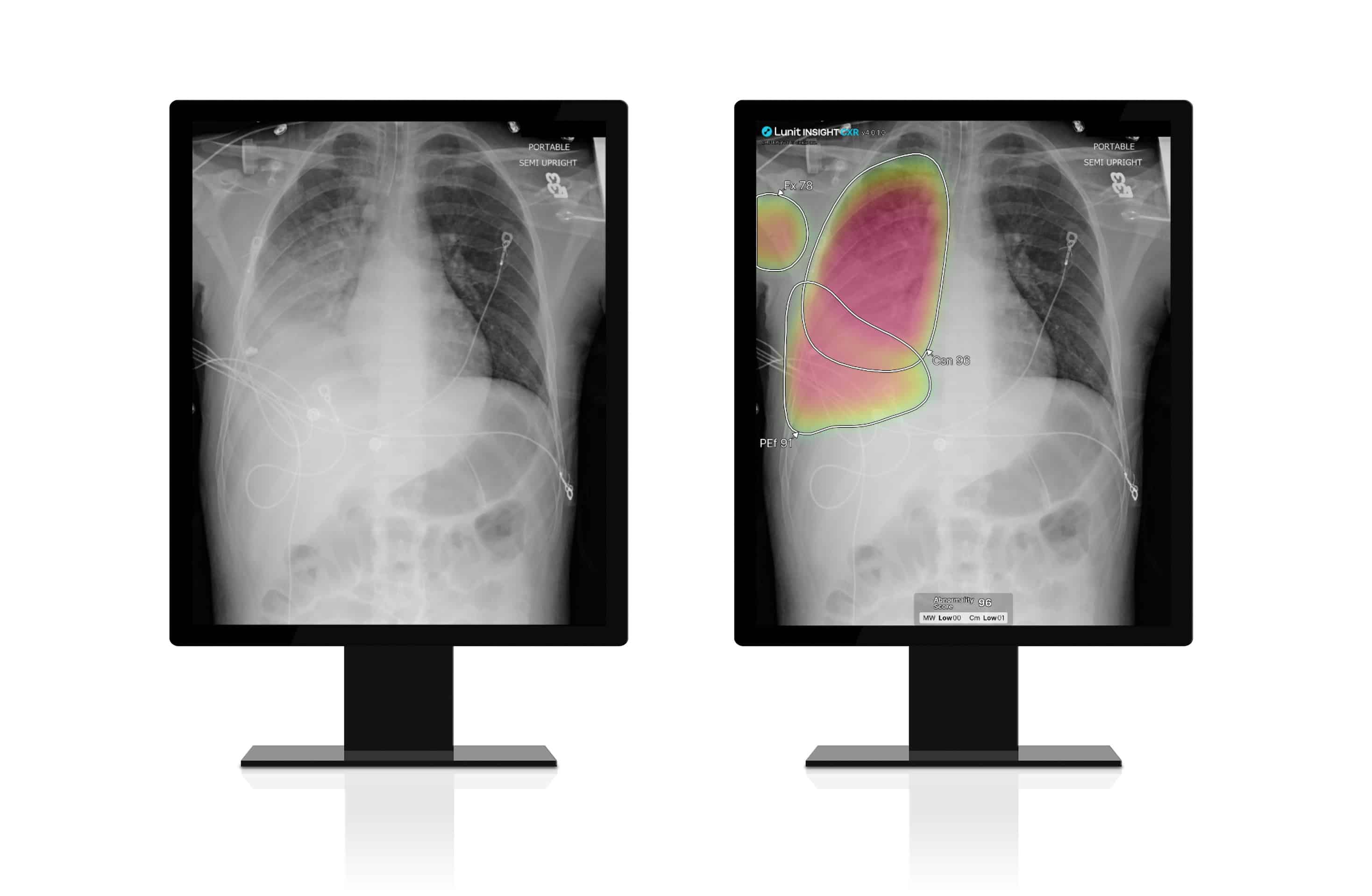
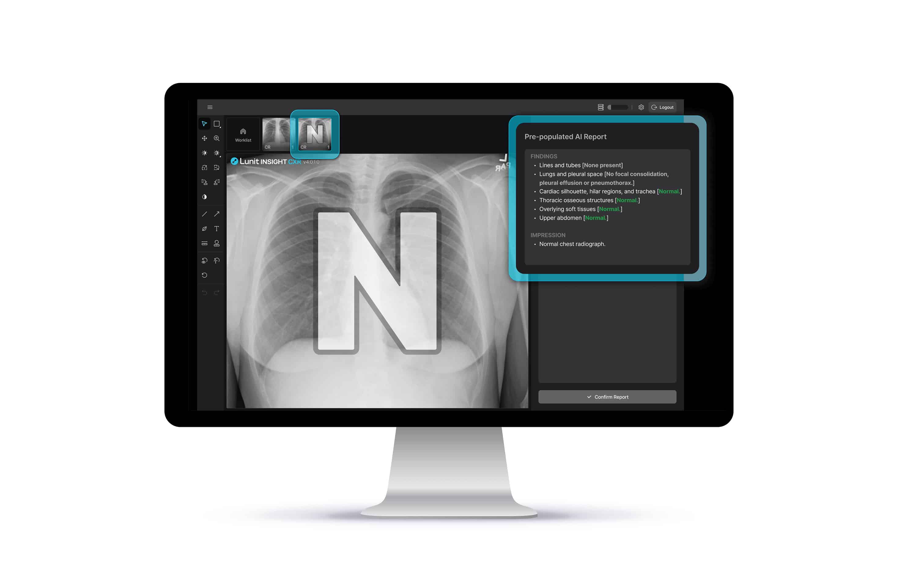
Built on very high sensitivity, Normal Flagging identifies high confidence normal cases–streamlining workflow and enhancing operational efficiency.
This allows radiologists to focus on abnormal and high‑risk cases, reallocate time to higher‑value imaging, and reduce repetitive workload and burnout.
Automatically surface changes in findings with current-prior comparison tools.
Including automated nodule progression and tracking for pneumothorax, pleural effusion, and consolidation.
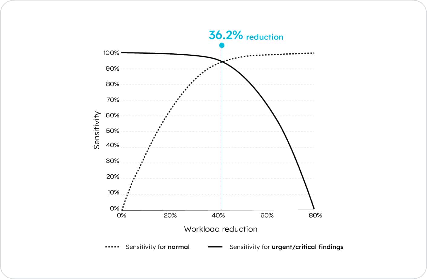
Lunit INSIGHT CXR helps radiologists manage high volumes by worklist management and surfacing critical cases. In studies*, it reduced reading workload by 36.2% while maintaining 95% sensitivity for urgent cases.¹
* This study was conducted based on CXR3.
1. Schalekamp et al, Performance of AI to Exclude Normal Chest Radiographs to Reduce Radiologists’ Workload. Published in European Radiology, 2024.
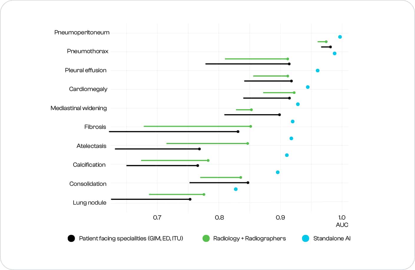
Whether read by radiologists or referring clinicians, Lunit INSIGHT CXR supports more accurate and efficient interpretations across departments.²
2. Shih A, et al. Does AI Assistance Improve Clinical Interpretation of Inpatient and Emergency Department Chest X-Rays? Abstract presented at RSNA 2024.
By monitoring the streaming of your data day-to-day, it helps you identify the best settings for your needs and optimize how Lunit INSIGHT supports your team.
*Please note: INSIGHT Manager is a non-medical device.
“Some cases are hard to analyze definitively, such as the ones with abnormalities hidden by blood vessels and rib.
Lunit AI helps me double-check these kinds of cases and confirm my analysis with certainty.”
“I sped up the interpretation of the chest radiology imaging studies and this allowed me to improve the relationship with the referring doctors from various hospital units, such as the Emergency Room, or Intensive Care.”

Stay informed with the latest research, product updates, and global partnerships driving innovation in AI-powered cancer screening.
Access product guides, validation briefs, and educational content to support evaluation and implementation of Lunit INSIGHT CXR.