Advanced feature analysis for TME biomarkers

Advanced feature analysis for TME biomarkers

Enable deeper understanding of tumor microenvironment and candidate biomarker signals through spatial and cell-level morphology analysis.
Tumor-agnostic platform	for H&E based CDx

Tumor-agnostic platform for H&E based CDx

Support broad application across drug development programs for H&E based AI-powered CDx regardless of cancer type.
Real world evidence

Real world evidence

Tested on 2,300+ real-world patient samples, with results published in JCO and JITC.

AI-Powered TME Insights

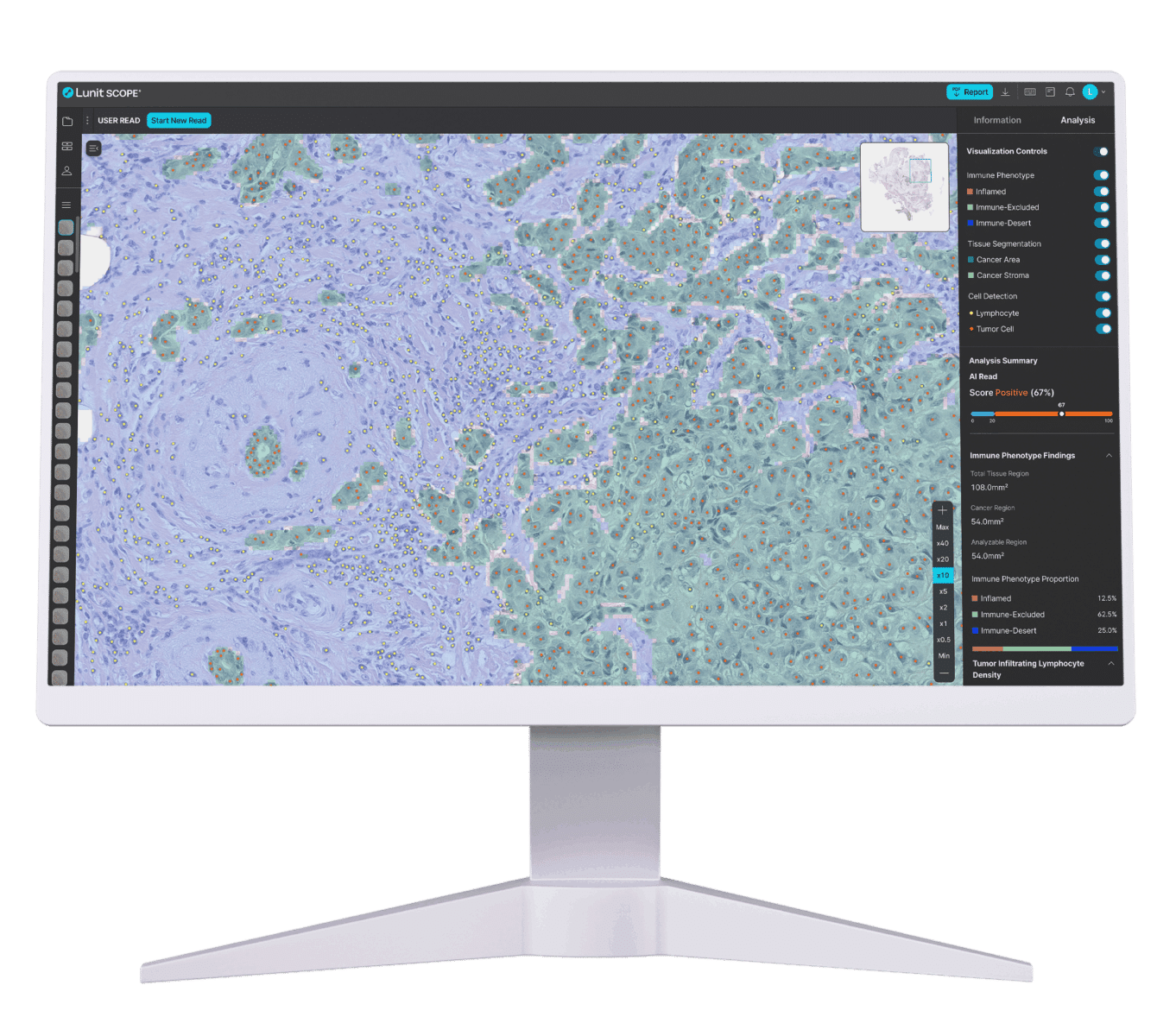
Lunit SCOPE IO® enables biopharma companies to identify tumor-agnostic, H&E-based immune biomarkers with precision and scale—accelerating biomarker development and enriching immunotherapy trials without additional stains or assays.

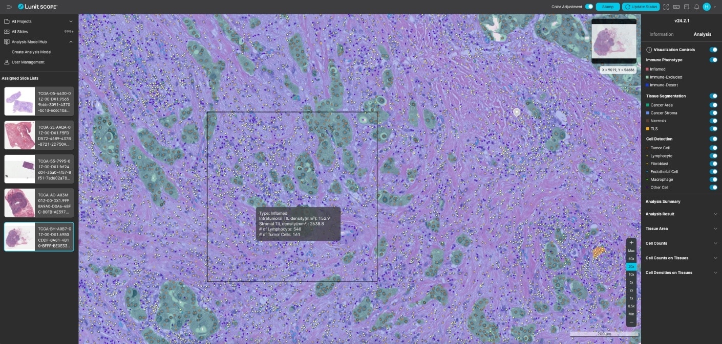
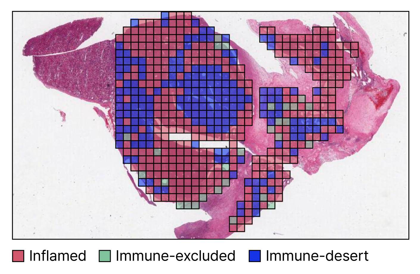
Trained on 50,000+ whole-slide images and over 10 million cell annotations by hundreds of pathologists worldwide, this AI-powered solution analyzes tumor microenvironments by performing quantitative immune phenotyping and assessing the immune landscape.

How Lunit SCOPE IO Supports Your Team

Digital pathology image showing AI-powered analysis of H&E tissue slides to identify tumor cells, fibroblasts, lymphocytes, and spatial patterns for biomarker discovery.

Feature identification for biomarker formation

Extract key morphological features from H&E slides—including TLS, necrosis, mitosis, tumor-stroma ratio, and spatial patterns and tumor-stroma borders—alongside relevant cell types, like tumor cells, lymphocytes, macrophages, fibroblasts, and endothelial cells. Analyze TILs and classify immune phenotypes (Inflamed, Excluded, and Desert) to support biomarker discovery.
Tissue sample with AI-generated colored grid showing immune phenotypes labeled as inflamed, immune-excluded, and immune-desert to support companion diagnostic development.

CDx-ready AI software

Leverage AI-powered H&E analysis across the clinical pathway for companion diagnostic development—from early-stage biomarker discovery to predicting response to immunotherapies that support patient selection.

Case Study: AI-powered Biomarker Development Using Lunit SCOPE IO

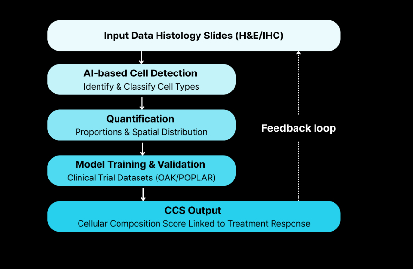
Lunit SCOPE IO enabled AI-driven histology analysis to quantify how the tumor microenvironment’s cellular composition correlates with response to atezolizumab. The AI-powered biomarker has shown to hold great potential for patient stratification and for informing treatment selection strategies.

Case Study: AI-powered Biomarker Development Using Lunit SCOPE IO

AI Pathology Software Built to Support Biopharma Innovation

Our digital image analysis software helps biopharma companies scale oncology R&D, advance biomarker strategies, and optimize trials. Lunit is built for end-to-end CDx development, with mature regulatory capabilities, broad scanner and file type compatibility, and scalable deployment across digital pathology platforms and CRO partners.
Breakthrough Insights to Drive Success in Drug Development

Breakthrough Insights to Drive Success in Drug Development

Equip your team with validated AI solutions that accelerate biomarker discovery, enhance translational strategies, and extract TME insights from H&E.

Frequently Asked Questions

What differentiates Lunit as a partner in AI-powered image analysis?

Our deep expertise, robust clinical evidence, and partnerships with many CROs and digital pathology platform providers set us apart. We prioritize collaboration and offer versatile solutions that are compatible with a wide range of scanners and IMS.