Trusted results

Trusted results

Trained with 1M+ cell annotations by 150+ pathologists worldwide to generate validated, reliable results.
Reduced discrepancy

Reduced discrepancy

Reduce interpretation discrepancies between readers with objective results.
Seamless interoperability

Seamless interoperability

Offers seamless interoperability with integration into platforms including Leica Biosystems' Aperio AI Store, Indica Labs' HALO AP®, and Roche navify®.

AI-Powered Detection and Classification of PD-L1 Expression

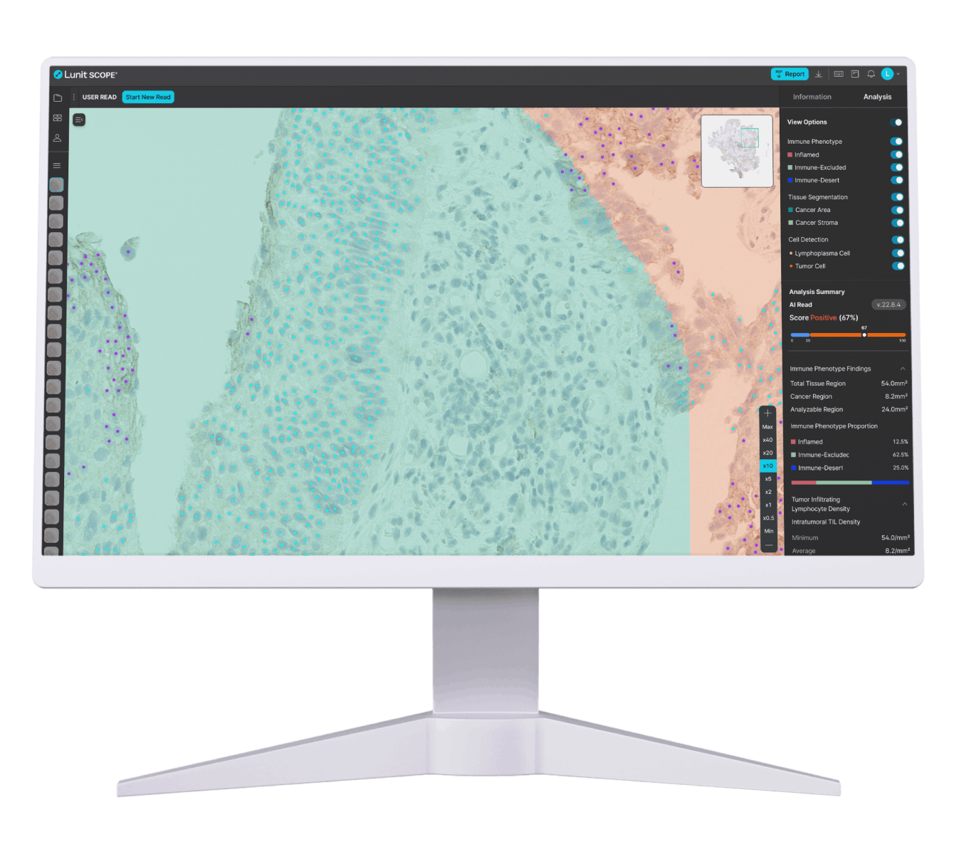
Lunit SCOPE PD-L1 assists in detecting and classifying PD-L1 biomarker expression by analyzing digitized IHC whole slide images from formalin-fixed paraffin embedded (FFPE) tissue.

It supports consistent, quantitative assessment of PD-L1 status and is designed to enhance productivity.

AI-Powered Detection and Classification of PD-L1 Expression

Proven AI-Powered Speed and Accuracy

Bar charts showing that AI-assisted PD-L1 analysis improved objective response rate from 2.3% to 15% and progression-free survival from 1.8 to 2.7 months in edge cases initially scored as negative.

Improves accuracy

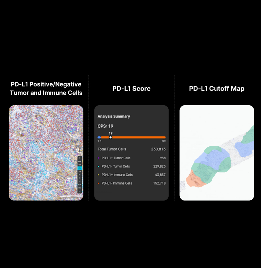
Lunit SCOPE PD-L1 enhances scoring accuracy for complex images, including edge or ambiguous samples1.

¹Kim H. et al. 2024. Clinical Validation of Artificial Intelligence–Powered PD-L1 Tumor Proportion Score Interpretation for Immune Checkpoint Inhibitor Response Prediction in Non–Small Cell Lung Cancer, JCO Precis Oncol, 2024;8: e2300556

Line chart showing PD-L1 scoring discrepancies among pathologists reduced from 32.1% to 10.3% with AI assistance, improving scoring agreement for tumor proportion scores below 1%.

Enhances scoring agreement

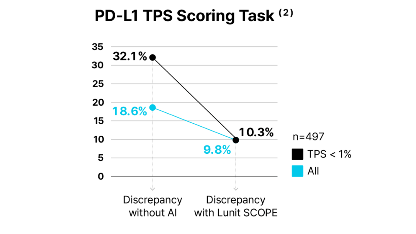
Lunit SCOPE PD-L1 delivers objective, reliable analysis results with proven support for reading consistency and improved scoring agreement.²

²Choi S. et al. 2022. AI–powered programmed death ligand 1 analyser reduces interobserver variation in tumour proportion score for non–small cell lung cancer with better prediction

Shortens reading time

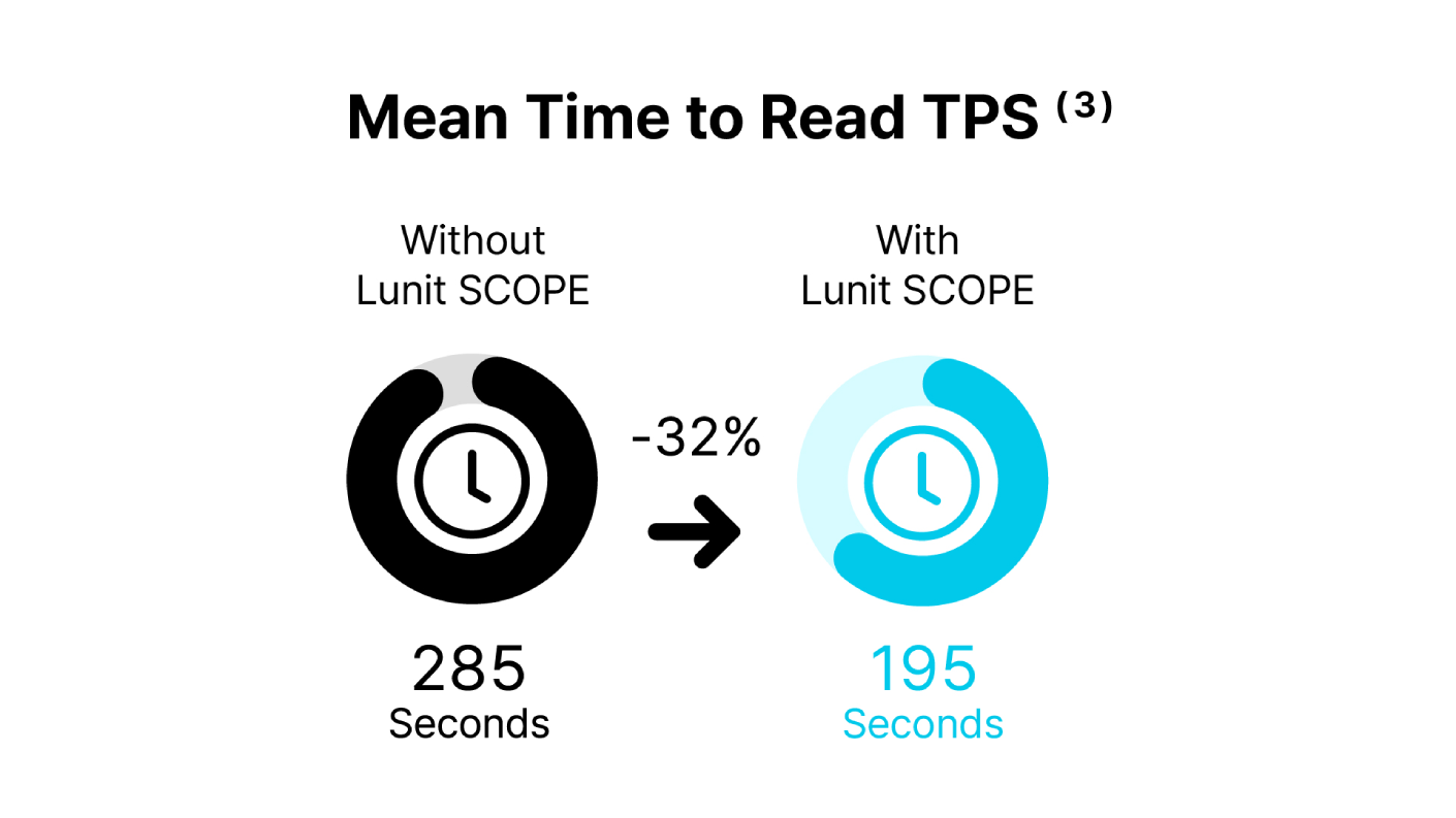
Lunit SCOPE PD-L1 can improve efficiency by reducing reading time by 32%.³

³Kim S. et al. 2022. Observer performance study to examine the feasibility of the AI-powered PD-L1 analyzer to assist pathologists’ assessment of PD-L1 expression using tumor proportion score in non–small cell lung cancer, JCO, 2022;40(16)

Infographic showing average PD-L1 reading time reduced by 32%, from 285 seconds without AI to 195 seconds with AI support.

AI Pathology Software Built to Support Accuracy and Productivity

Lunit SCOPE PD-L1 is integrated into digital pathology platforms, including Leica Biosystems' Aperio AI Store, Indica Labs' HALO AP®, and Roche navify®. It empowers research and pharma worldwide with ease of use, seamless operability, powerful analytics, and unmatched processing speeds in digital pathology—enabling quantitative tissue analysis with precision and efficiency.
Boost Your Efficiency with Fast, Accurate Insights

Boost Your Efficiency with Fast, Accurate Insights

Equip your team with PD-L1 quantification software that improves accuracy, reduces inter-observer variability, and improves workflow efficiency.

Frequently Asked Questions

What differentiates you as a partner in AI-powered image analysis?

Our deep expertise, robust clinical evidence, and partnerships with many CROs and digital pathology platform providers set us apart. We prioritize collaboration and offer versatile solutions that are compatible with a wide range of scanners and IMS.