- Background
- Product Description
- Major Benefits
- Example Cases
Background
What if the most basic test can catch
the least visible findings?
When Detected
Early by AI*5-year survival
*Reference: AJCC 8th Edition
Product Description
Lunit INSIGHT CXR covers
the vast majority of findings.
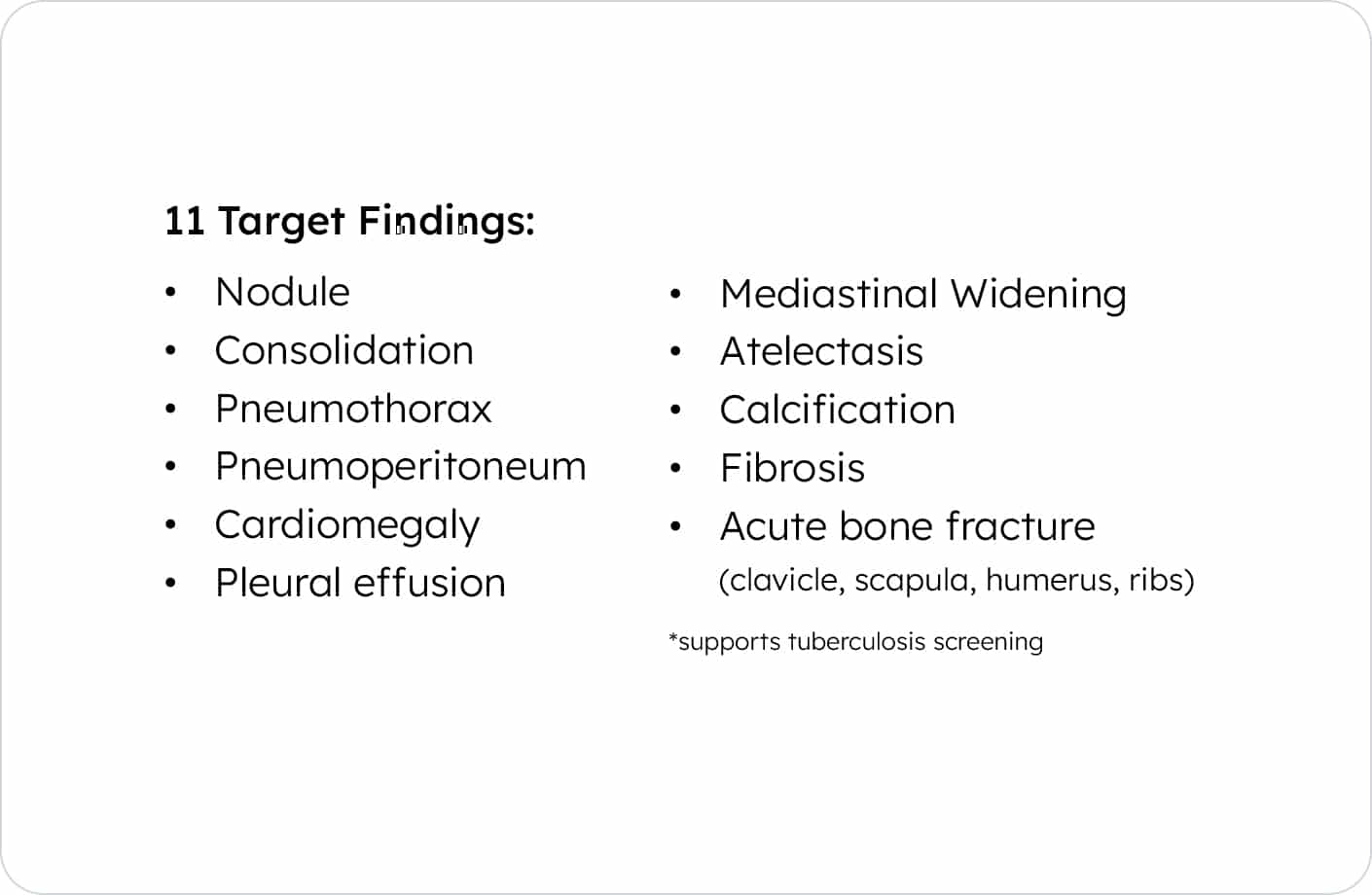
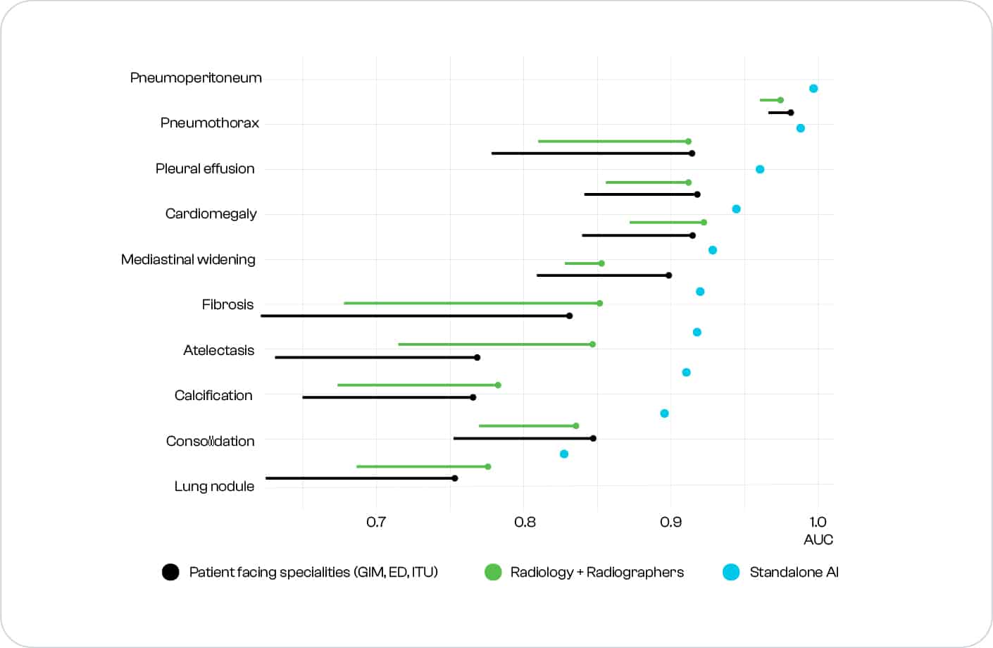
Accurately detects 10 of the most common findings in
a chest x-ray.Developed by using Lunit’s cutting-edge
deep learning technology.*Supports Tuberculosis Screening
Generated by
Lunit INSIGHT CXRDetected Location
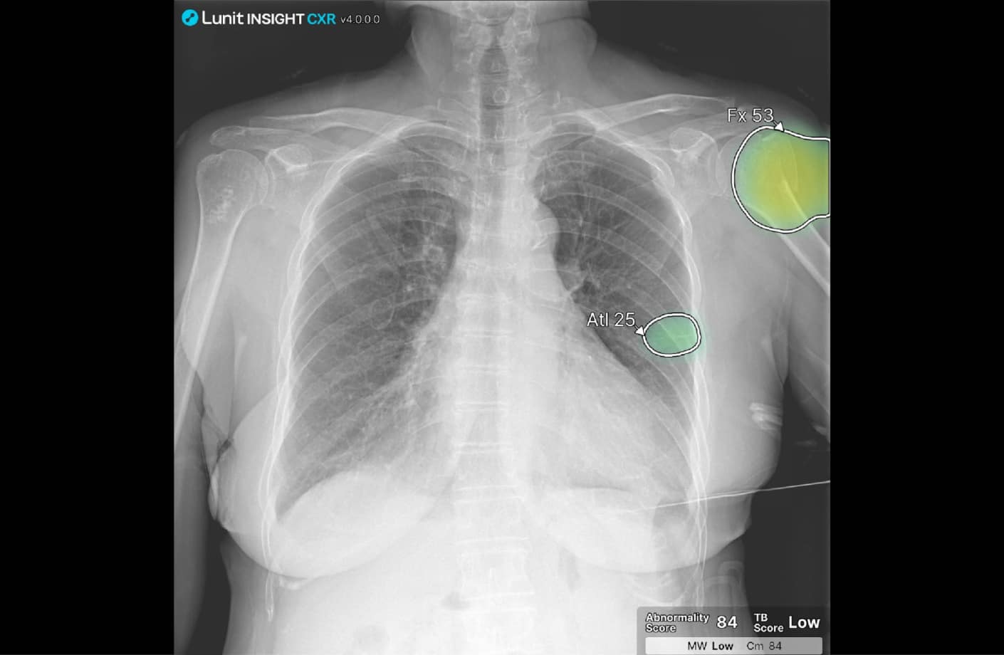
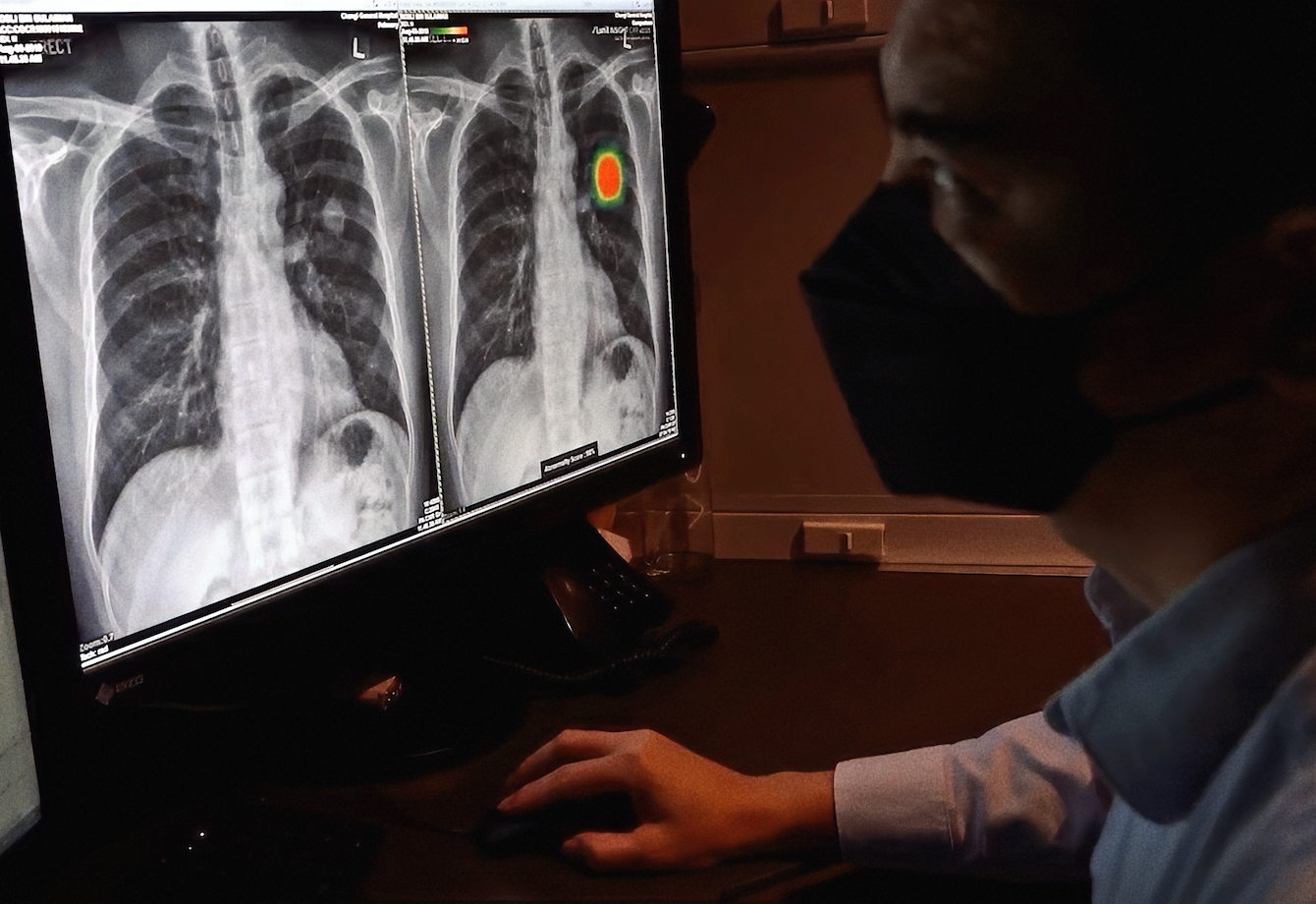
The AI generates the location information of detected lesions in the form of heatmaps and outlines.
Abnormality Score
The AI generates an abnormality score which reflects the AI’s calculation of the actual presence of the detected lesion.
AI Report
The AI provides a “case report” that summarizes the overall analysis result, narrowed down to each finding.
Major Benefits
Accurate and efficient diagnosis
boosted with AI
Lunit INSIGHT in Yonsei Severance Hospital,
Yongin, KoreaFast triage of
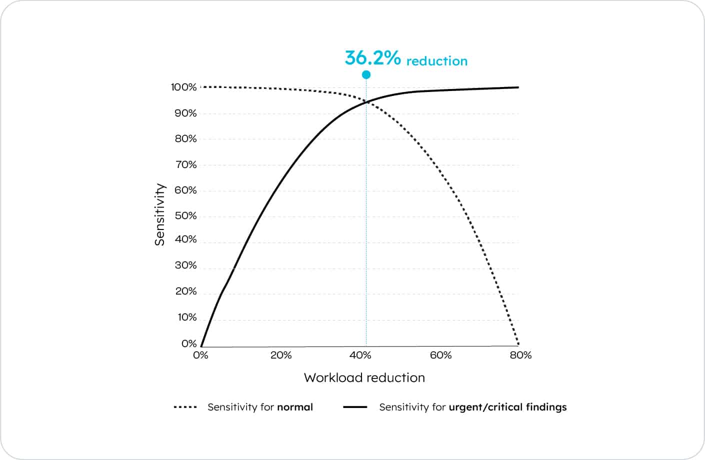
normal casesTriage normal cases quickly and focus on reading abnormal cases
Efficient reading via
exam prioritizationPrioritize cases according to each abnormality scores, reducing reading time by 65% for normal cases and 25% for abnormal cases.1
Early diagnosis of
lung cancerReduce false negative cases and detect lung cancer at early stages with AI-aided detection of small, subtle pulmonary nodules.9
Example Cases
Never miss a finding.
A nodule, diagnosed as lung cancer, hidden behind the heart is properly detected, with an abnormality score of 44%. This case was missed by 8 out of 15 radiologists.
Abnormality Score
44
%Radiologists Missed
8 out of 15
A nodule, diagnosed as lung cancer, in the right upper lung field is properly detected, with an abnormality score of 66%. This case was missed by 9 out of 9 radiologists.
Abnormality Score
66
%Radiologists Missed
9 out of 9
A nodule, diagnosed as lung cancer, hidden behind the diaphragm is properly detected, with an abnormality score of 96%. This case was missed by 5 out of 9 radiologists.
Abnormality Score
96
%Radiologists Missed
5 out of 9
Distributed in
partnership withMajor Customers
Publications featuring
Lunit INSIGHT CXR
Our solutions


Watch and learn more about
Lunit INSIGHT CXR
Lunit YouTubeReference
- Background
1Radiation UNSCotEoA. Sources and effects of ionizing radiation: sources: United Nations Publications; 2000.
2Quekel LG, Kessels AG, Goei R, van Engelshoven JM. Miss rate of lung cancer on the chest radiograph in clinical practice. Chest 1999;115:720-4.
3Forrest JV, Friedman PJ. Radiologic errors in patients with lung cancer. Wes J Med 1981;134:485.
4Kesselman A, Soroosh G, Mollura DJ, et al. 2015 RAD-AID Conference on International Radiology for Developing Countries: the evolving global radiology landscape. J Am Coll Radiol 2016;13:1139-44.
5Monnier-Cholley L, Carrat F, Cholley BP, Tubiana J-M, Arrivé L. Detection of lung cancer on radiographs: receiver operating characteristic analyses of radiologists’, pulmonologists’, and anesthesiologists’ performance. Radiology 2004;233:799-805.
6Eng J, Mysko WK, Weller GE, et al. Interpretation of emergency department radiographs: a comparison of emergency medicine physicians with radiologists, residents with faculty, and film with digital display. Am J Roentgenol 2000;175:1233-8.
7Potchen EJ, Cooper TG, Sierra AE, et al. Measuring performance in chest radiography. Radiology 2000;217:456-9.
8Milner RC, Culpan G, Snaith B.Radiographer reporting in the UK: is the current scope of practice limiting plain-film reporting capacity? Br J Radiol. 2016 Sep 89(1065):20160228. doi:
- Major Benefits
1Internal test results
2Ju Gang Nam, Sunggyun Park, et al., Development and Validation of Deep Learning–based Automatic Detection Algorithm for Malignant Pulmonary Nodules on Chest Radiographs, Radiology, 2018
3Eui Jin Hwang, Sunggyun Park, et al., Development and Validation of a Deep Learning-based Automatic Detection Algorithm for Active Pulmonary Tuberculosis on Chest Radiographs, Clinical Infectious Diseases, 2018
4Eui Jin Hwang, Sunggyun Park, Kwang-Nam Jin, et al., Development and Validation of a Deep Learning–Based Automated Detection Algorithm for Major Thoracic Diseases on Chest Radiographs, JAMA Network Open, 2019
5Jong Hyuk Lee, Sunggyun Park, et al., Deep learning–based automated detection algorithm for active pulmonary tuberculosis on chest radiographs: diagnostic performance in systematic screening of asymptomatic individuals, European Radiology, 2020
6Eui Jin Hwang, Jung Hee Hong, et al., Deep learning algorithm for surveillance of pneumothorax after lung biopsy: a multicenter diagnostic cohort study, European Radiology, 2020
7Jong Hyuk Lee, Hye Young Sun, et al., Performance of a Deep Learning Algorithm Compared with Radiologic Interpretation for Lung Cancer Detection on Chest Radiographs in a Health Screening Population, Radiology, 2020
8Hyunsuk Yoo, Ki Hwan Kim, et al., Validation of a Deep Learning Algorithm for the Detection of Malignant Pulmonary Nodules in Chest Radiographs, JAMA Network Open. 2020
9Sowon Jang, Hwayoung Song, et al., Deep Learning–based Automatic Detection Algorithm for Reducing Overlooked Lung Cancers on Chest Radiographs, Radiology, 2020
10Jae Hyun Kim, Jin Young Kim, et al., Clinical Validation of a Deep Learning Algorithm for Detection of Pneumonia on Chest Radiographs in Emergency Department Patients with Acute Febrile Respiratory Illness, Journal of Clinical Medicine, 2020